Rv Lv Ratio Echo
Https Encrypted Tbn0 Gstatic Com Images Q Tbn 3aand9gctrtldus7mggg4dy8o1fwklwf3yeeeu 8qcng Usqp Cau
Https Edus Ucsf Edu Sites Edus Ucsf Edu Files Wysiwyg Dresden 20rv 20dilation 20pe Pdf
Basic Haemodynamic Assessment With Echo Iheartscan
Differentiating Acute Versus Chronic Right Heart Failure With Bedside Echocardiography Emra
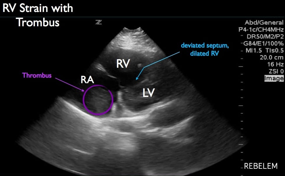
Diagnosis Of Right Ventricular Strain With Transthoracic Echocardiography Rebel Em Emergency Medicine Blog

Imaging Right Left Ventricular Interactions Jacc Cardiovascular Imaging

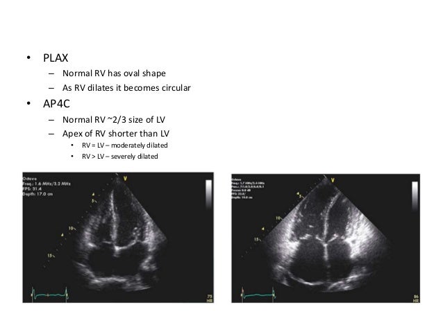
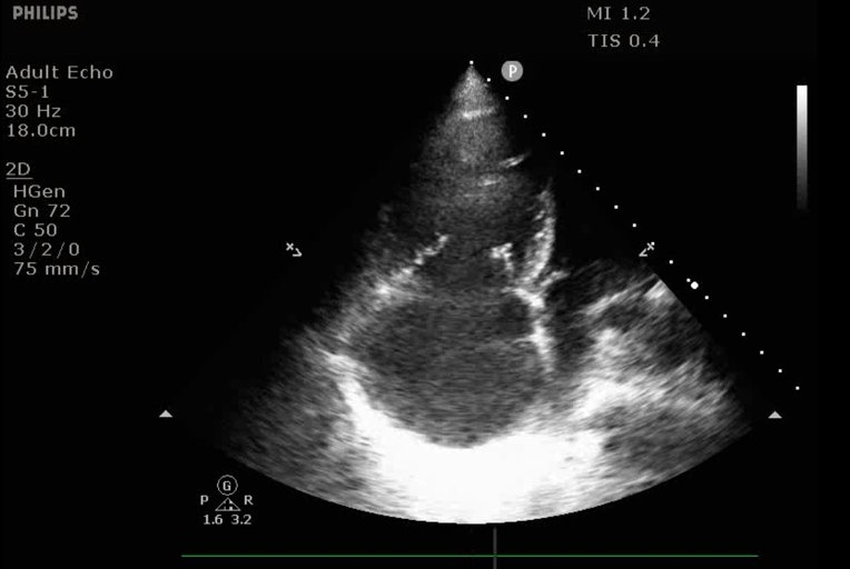
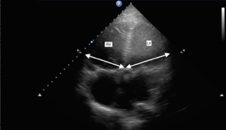
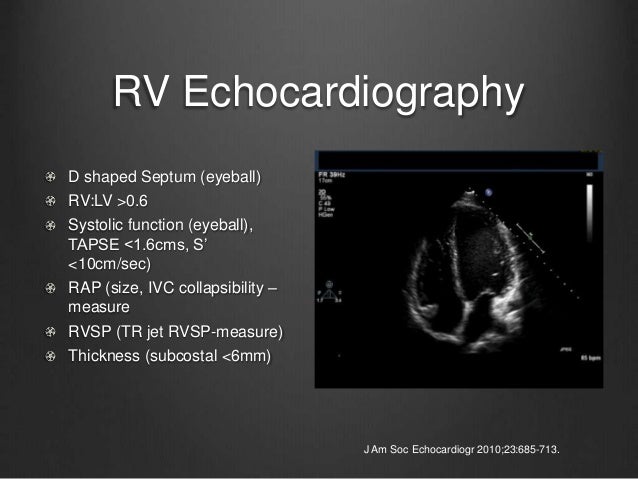
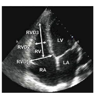
The assessment of rv function starts with the measurement of rv dimentions and the qualitative evaluation of its function.
Rv lv ratio echo.

Apical Four Chamber View Panel A Demonstrates Normal Rv Lv Size Ratio Download Scientific Diagram

Acep American College Of Emergency Physicians
Ctpa Demonstrating The Rv Lv Ratio Measurement Note Ctpa Computed Download Scientific Diagram
Submassive Pe Emory School Of Medicine
Echocardiography In Pulmonary Embolism Ppt Video Online Download

Figure 1 From Increased Right To Left Ventricle Diameter Ratio Is A Strong Predictor Of Right Ventricular Failure After Left Ventricular Assist Device Semantic Scholar
Https Www Jhltonline Org Article S1053 2498 16 00013 9 Pdf
Https Encrypted Tbn0 Gstatic Com Images Q Tbn 3aand9gcryyxpe I0f Apg9ghlpraeedhflrw1vi16uq Usqp Cau
A Practical Approach To Goal Directed Echocardiography In The Critical Care Setting Springerlink
Icn Victoria Burrell On Rv Failure For The Intensivist
3 3 2 Right Ventricular Size 123sonography

Constrictive Pericarditis Role Of Echocardiography And Magnetic Resonance Imaging

Allied Health Allied Health Testing Diagnostics Echocardiogram

Pdf Echocardiography In Pediatric Pulmonary Hypertension

Colour Doppler Echo In Tetralogy Of Fallot Youtube Tetralogy Echo Echocardiogram
Https Criticalcarecanada Com Presentations 2016 Peep And The Rv During Mechanical Ventilation Pdf

Hypertrophic Cardiomyopathy Hypertrophic Cardiomyopathy Medical School Stuff Heart Conditions

Rv Versus Lv Youtube
Https Encrypted Tbn0 Gstatic Com Images Q Tbn 3aand9gcra85szsxx3hpakprypgckbrujloz3yo Uzmxoqw8g L5raoawh Usqp Cau

Rvot View Tee Cardiac Sonography Ultrasound Cardiology

Qp Qs Ratio In Echo Echocardiography Barnard Health Care In 2020 Echocardiogram Ductus Arteriosus Ventricular Septal Defect
Emdocs Net Emergency Medicine Educationus Probe When Does An Effusion Become Pericardial Tamponade Emdocs Net Emergency Medicine Education

Echocardiographic Predictors Of Pulmonary Embolism In Patients Referred For Helical Ct Lodato 2008 Echocardiography Wiley Online Library

Presence And Prognostic Value Of Ventricular Diastolic Function In Arrhythmogenic Right Ventricular Cardiomyopathy Sadeghpour Echocardiography Wiley Online Library
Source : pinterest.com
